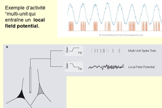
Because neurons are so small and numerous and there are so many different types of electrical activity on dendrites, cell body and axon, interpreting what is registered in the brain presents enormous challenges. An electrode can be lowered into the extracellular medium between neurons and thus capture the sum of the electrical activities generated by various electrochemical phenomena in the many neurons around its tip.
This signal in English is called as ” local field capabilities », or LFP. By comparing this activity to a reference point, its differences can be measured in millivolts. Typically, we would then record a few thousand neurons within a radius of 100 to 300 micrometers, sometimes as small as 1 millimeter, and it is still a matter of debate. This global signal can then be filtered to isolate oscillating phenomena in very specific frequency windows. For example, what we pick up at the signal’s highest frequencies, from 600 to 3,000 Hz, corresponds to a superposition of action potentials emitted by a few thousand neurons. around 250 Hz, then we filter the LFPs. For frequencies below 200 Hz, it will be a reflection of inputs that reach the dendrites of neurons adjacent to the electrode tip in a wider radius.
Then we get to scale Electrocortical diagramThis means thatApply a larger electrode directly to the surface of the cortex. Therefore, here it is also necessary to open the skull, which makes it an invasive technique of little use in humans except in cases where patients are waiting for brain surgery to remove a tumor or a focus of epilepsy, in which the skull is opened. anyway necessary. Then we pick up More neurons activity within a radius of about 3 millimeters around the electrode. The next level is the level ElectroencephalogramEEG, or EEG, which records the overall electrical activity of the brain by attaching small electrodes to the scalp.
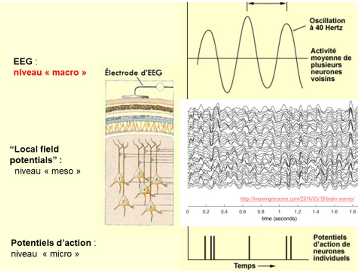
This non-invasive technique was developed in the mid-1920s by the German Hans Berger. He was able to record such weak currents through the meninges, skull and scalp because the apical dendrites of pyramidal neurons in the cortex all signal to the surface and the synchronous activity occurring at the same time is strong enough that he could detect it on the surface of the skull by greatly amplifying it. Unlike whole brain imaging techniques such as Pet check up or fMRI, EEG is a direct measurement of the electrical activity of neurons with a time resolution of milliseconds. On the other hand, as voltage decreases with the square of distance, activity in subcortical structures is difficult to detect using an EEG and spatial resolution is not its strength. Rather, it is the ability to pick up with great accuracy when certain events occur in the brain. To draw an analogy with the people out there in the stands watching a baseball game, we are quite unable to discern who is talking to whom at the moment, but if a home run were run we would at that very moment hear cheers from the crowd.
In slightly the same way, since the 1970s, we have started recording EEGs while presenting all kinds of stimuli to the subject. It is run on an EEG to track the so-called raised potential. For example, when the last word of a sentence is abnormal, the EEG shows a negative skew about 400 ms after the subject has finished reading the sentence.
Finally, since the EEG records the electrical activity of the brain, the MagnetoencephalographyMEG, or MEG, measures the magnetic field associated with this electric current through magnetic field-sensitive detectors lined up around the head. MEG is the safest of the various brain imaging technologies because the device does not send anything into the brain and does not touch the head.
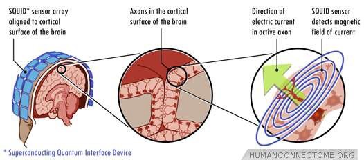
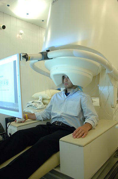 The kind of big hair dryer we put our head under contains liquid helium at -260°C which cools magnetometers based on superconductors that then become extremely sensitive to the weak magnetic fields produced by neurons when cooled to these temperatures. The advantage of measuring these magnetic fields, even if they are very weak, is that they pass through the skull and other tissues without any distortion, unlike the EEG whose signals are more distorted. The temporal sampling rate of MEG is similar to that of EEG at about 1,000 samples per second It has a fairly good spatial resolution of the order of a few millimeters. And Technology continues to improve…
freshidea-AdobeStock-ef12.jpeg)